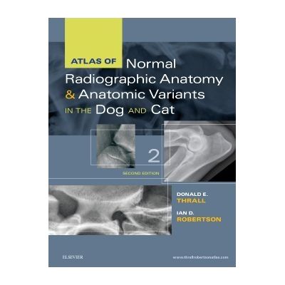
Atlas of Normal Radiographic Anatomy and Anatomic Variants in the Dog and Cat - Donald H. Thrall

Detalii Atlas of Normal Radiographic Anatomy
Atlas of Normal Radiographic Anatomy - Disponibil la grupdzc.ro
Pe YEO găsești Atlas of Normal Radiographic Anatomy de la Saunders, în categoria Carti medicale.
Indiferent de nevoile tale, Atlas of Normal Radiographic Anatomy and Anatomic Variants in the Dog and Cat - Donald H. Thrall din categoria Carti medicale îți poate aduce un echilibru perfect între calitate și preț, cu avantaje practice și moderne.
Preț: 703.5 Lei
Caracteristicile produsului Atlas of Normal Radiographic Anatomy
- Brand: Saunders
- Categoria: Carti medicale
- Magazin: grupdzc.ro
- Ultima actualizare: 06-11-2024 01:21:16
Comandă Atlas of Normal Radiographic Anatomy Online, Simplu și Rapid
Prin intermediul platformei YEO, poți comanda Atlas of Normal Radiographic Anatomy de la grupdzc.ro rapid și în siguranță. Bucură-te de o experiență de cumpărături online optimizată și descoperă cele mai bune oferte actualizate constant.
Descriere magazin:
Equip yourself to make accurate diagnoses and achieve successful treatment outcomes with this highly visual comprehensive atlas. Featuring a substantial number of new high contrast images Atlas of Normal Radiographic Anatomy and Anatomic Variants in the Dog and Cat 2nd Edition provides an in-depth look at both normal and non-standard subjects along with demonstrations of proper technique and image interpretations. Expert authors Donald E. Thrall and Ian D. Robertson describe a wider range of normal as compared to competing books - not only showing standard dogs and cats but also non-standard subjects such as overweight and underweight pets and animals with breed-specific variations. Every body part is put into context with a textual description to help explain why a structure appears as it does in radiographs and enabling practitioners to appreciate variations of normal that are not included based on an understanding of basic radiographic principles. Key Features Radiographic images of normal or standard prototypical animals are supplemented by images of non-standard subjects exhibiting breed-specific differences physiologic variants or common congenital malformations. Images that depict a wider range of normal - such as images that detail the natural growth and aging characteristics of normal pediatric and senior animals - prevents clinical under- and over-diagnosing. In-depth coverage of patient positioning and radiographic exposure guidelines assist clinicians in producing the very best results. Unlabeled radiographs along side labeled counterparts clarifies important anatomic structures of clinical interest. High-quality digital images provide excellent contrast resolution and better visibility of normal structures to assist clinicians in making accurate diagnoses. Brief descriptive text and explanatory legends accompany all images to help put concepts into the proper context. An overview of radiographic technique includes the effects of patient positioning respiration and exposure factors. New to this Edition NEW Companion website features additional radiographic CT scans and more than 100 questions with answers and rationales. NEW Additional CT and 3D images have been added to each chapter to help clinicians better evaluate the detail of bony structures. NEW Breed-specific images of dogs and cats are included throughout the atlas to help clinicians better understand the variances in different breeds. NEW Updated material on oblique view radiography provides a better understanding of an alternative approach to radiography particularly in fracture cases. NEW 8. 5 x 11 trim size makes the atlas easy to store.

Produse asemănătoare

Atlas of Normal Radiographic Anatomy and Anatomic Variants in the Dog and Cat - Donald H. Thrall
![]() grupdzc.ro
grupdzc.ro
Actualizat in 06/11/2024
703.5 Lei
Produse marca Saunders

Success in Practical/Vocational Nursing: From Student to Leader, Paperback/Patricia Knecht
![]() elefant.ro
elefant.ro
Actualizat in 20/12/2024
367.99 Lei

Understanding Nursing Research: Building an Evidence-Based Practice, Paperback/Susan K. Grove
![]() elefant.ro
elefant.ro
Actualizat in 20/12/2024
634.99 Lei

Mastering Neuroscience: A Laboratory Guide, Paperback/Roseann Cianciulli Schaaf
![]() elefant.ro
elefant.ro
Actualizat in 20/12/2024
322.99 Lei

Veterinary Dentistry: A Team Approach, Paperback/Steven E. Holmstrom
![]() elefant.ro
elefant.ro
Actualizat in 20/12/2024
508.99 Lei

Atlas of Dental Radiography in Dogs and Cats, Hardcover/Gregg A. DuPont
![]() elefant.ro
elefant.ro
Actualizat in 20/12/2024
647.99 Lei
